Grand Rounds
Tuesday, November 6th 2018
Topic: "MR Lymphangiography and Lymphedema"

Jeffrey Maki, MD, PhD
Visiting Professor of Radiology
Section Chief, Abdominal Imaging
University of Colorado, School of Medicine
Wednesday, October 24th 2018
Topic: "Endovascular Therapy For Clinical Limb Ischemia"
Robert Lookstein, MD MHCDL, FSIR FAHA FSVM
Vice Chairman, Interventional Services
Professor of Radiology and Surgery
Mt. Sinai Health System
Monday, October 8, 2018
Topic: "Clinical and Practical Applications of fMRI"
Andrei I. Holodny, MD, PhD (hon), FACR
Chief of the Neuroradiology
Service Director of the Functional MRI Laboratory
Department of Radiology
Memorial Sloan-Kettering Cancer Center
RSNA Travel Award
A winner for the RSNA Student Travel Award for young investigators is Hailiang Huang, MS, Breast Structural Noise Analysis of Narrow- and Wide-angle Tomosynthesis and Masking Risk Assessment for Mass Detection, who works in Professor Wei Zhao's Lab. 
Up to 430 of the top-rated abstracts received a $500 stipend to attend the 2018 Annual Meeting. Scientific abstracts from current RSNA member who are presently enrolled in a full time undergraduate or graduate program; clinical trainees who are currently enrolled in a full-time clinical training program; or postdoctoral trainees who were awarded a doctorate or equivalent degree no more than three years ago, were eligible for the award.
AAPM Award
Adrian Howansky, a Ph.D. candidate in Prof. Wei Zhao’s lab, took 2nd place in the John. R. Cameron Young Investigator symposium at this year's American Association of Physicists in Medicine (AAPM) Annual Meeting. The meeting was held from 7/29 to 8/2 in Nashville, Tennessee. Adrian presented a scientific paper entitled: “Guiding novel flat-panel detector designs with direct measurements of depth-dependent scintillator gain and blur”. This paper described experimental methods for determining the fundamental limits to the imaging properties of x-ray scintillators. Results of these experiments were applied to propose novel flat panel detector designs with superior image quality and dose efficiency than current detectors.

The paper was among 10 selected out of 321 submissions to enter the final competition symposium.
Grand Rounds
Tuesday, September 18th, 2018
TOPIC: “Leading Improvement Teams”
Lane F. Donnelly, MD
Chief Quality Officer
Lucile Packard Children’s Hospital at Stanford Professor
Associate Dean, Maternal and Child Health (Quality and Safety)
Stanford University School of Medicine
Monday, September 17th, 2018
TOPIC: "A Day in the Vascular Lab"
John S. Pellerito, MD, FACR, FAIUM, FSRU
Vice Chair, Education Department of Radiology
Zucker School of Medicine at Hofstra/Northwell North Shore University Hospital
Tuesday, September 11th, 2018
TOPIC: “Intraprocedural imaging and navigation tools in hepatobiliary interventions"
Baljendra S. Kapoor, MD FSIR FCIRSE
Associate Professor Vascular & Interventional Radiology Specialist
Cleveland Clinic Lerner College of Medicine of Case
Major Event
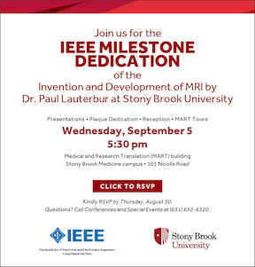 IEEE Milestones Dedication of Dr. Paul Lauterbur's Invention of MRI
September 5th, 2018 5:30pm MART Building
Click here to learn about Paul Lauterbur's, PhD, discovery using Nuclear Magnetic Resonance (NMR)
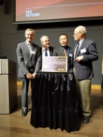
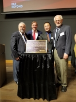
Unveiling of the IEEE Milestones Plaque, Sept 5, 2018
Left to right: Ken Kaushansky (SBU SOM Dean), Nick Golas (Long Island IEEE president), Tim Duong (Professor, Radiology VC for Research), Tom Coughlin (IEEE President-Elect)
Left to right: Nick Golas (Long Island IEEE president), Sam Stanley (President SBU), Tim Duong (Professor, Radiology VC for Research), Tom Coughlin (IEEE President-Elect)
Aug 2018 SBU Radiology summer research presentations
To see a list presentations by high school, undergraduate, and medical students please click the following, 2018summerreseaerch
AAPM showcases the ‘best in physics’
The “Best-in-Physics” session at the AAPM Annual Meeting showcase the studies that score highest in the abstract review process and are judged to reflect the highest level of scientific quality and innovation. The ever-popular electronic poster session includes presentations in therapy, imaging and joint imaging-therapy categories. Here’s our selection from this year’s chosen papers
SELENIUM RAMPS TOF-PET PERFORMANCE
In the imaging category, Andrew LaBella from SUNY Stony Brook University described NEW -HARP, an amorphous selenium photodetector for time-of-flight (TOF) PET. The aim of NEW-HARP (nano-electrode multi-well high-gain avalanche rushing photoconductor) is to achieve a sufficient time resolution to perform TOF-PET in simultaneous PET/MRI.For more information, please click here.
Grant awards (Aug 2018)
Two Bahl Endowment IDEA grants to Radiology faculty and colleagues:• Dr. Anat Biegon, PhD, received a grant to study aromatase role in lung cancer.
• Dr. Tim Duong, PhD and colleagues Drs. Parker, Hogan, Preston, Stefancin, Cahaney receive a grant to study pediatric chemo brain.
Grant award (Jul 2018) A team of oncologists, radiologists, and imaging scientists was awarded a grant over two years to develop novel imaging methods to study breast cancer and axillary lymph nodes.
Investigators: Cohen, Baer, Bernstein, Franceschi, Fisher, Duong
Grant award (Jul 2018) A team of imaging scientists, radiologists, oncologists were awarded a grant to study cognitive dysfunction in pediatric chemotherapy patients
Investigators: Duong, Parker, Hogan, Meyer, Preston, Cahaney, Stefancin
Grant award (2018) Dr. Chuan Huang, PhD, received a grant from Walk-for-Beauty foundation to study breast cancer

MIRA Lab publishes six Journal papers
Dr. Chuan Huang's MIRA Lab has published six Journal papers so far in 2018
T. Fukuda, M. Huang, A. Janardhanan, M.E. Schweitzer, C. Huang. Correlation of bone marrow cellularity and metabolic activity in healthy volunteers with simultaneous PET/MR imaging. In Press, Skeletal Radiology
K. Spuhler, J. Gardus III, Y. Gao, C. DeLorenzo, R. Parsey, C. Huang. Synthesis of patient-specific transmission data for PET/MRI attenuation correction in the brain using a convolutional neural network. In Press, Journal of Nuclear Medicine
Y. Zhao, M. Huang, J. Dingy, X. Zhang, K. Spuhler, S. Hu, M. Li, W. Fan, L Chen, X. Zhang, S. Li, Q Zhou, C. Huang. Prediction of abnormal bone density and osteoporosis from lumbar spine MR using modified Dixon Quant in 257 subjects with QCT as reference. In Press, Journal of Magnetic Resonance Imaging
C. Liu, J. Ding, K. Spuhler, Y. Gao, M. Serrano Sosa, M.A. Moriarty, S. Hussain, X. He, C. Liang, C. Huang. Preoperative prediction of sentinel lymph node metastasis in breast cancer by radiomic signatures from dynamic contrast-enhanced MRI. In Press, Journal of Magnetic Resonance Imaging
J. Dingy, A. Stopeck, Y. Gao, M.T. Marron, B.C. Wertheim, M.I. Altbach, J.P. Galons, D. Roe, F. Wang, G. Maskarinec, C. Thomson, P.A. Thompson, C. Huang. Reproducible automated breast density measure with no ionizing radiation using fat-water decomposition MRI. Early View, Journal of Magnetic Resonance Imaging
E. Bartlett, C. DeLorenzo, R. Parsey, C. Huang. Radio-Frequency Noise Contamination from PET Blood Sampling Pump: Effects on Structural MRI Image Quality in Simultaneous PET/MR Studies. Medical Physics 2018; 45(2)

Annonucements: Preclinical MRI Center
May 2018
Preclinical MRI Center is fully operational. We strive to provide in vivo imaging support for biomedical research at SBU.
We encourage a wide range of collaborations between various groups to explore new imaging techniques,
novel imaging contrast agents, or new applications in various research fields. It houses two state-of-the-art animal scanners:
- Bruker 7.0 Tesla MRI
- Bruker 9.4 Tesla MRI
- Computational infrastructure to assist image analysis
- High-resolution PET insert for simultaneous MRI-PET study
- Supportive infrastructure
Preclinical MRI Center underwrites all development costs for promising and fundable projects and helps analzye data
For limited time, SOM provides matching fund to support pilot/feasibility studies @ little to no costs to investigators
For limited time, SB Cancer Center provides matching fund to support pilot/feasibility studies @ little to no costs to investigators
Click here or contact Dr. Tim Duong, PhD (Director) more information
Noteable 2018 Summer Research Scholars
May 2018
SBM Radiology welcomes the following
Scholarly Concentration Program awardees: James Kang, Danling Chen, Shauheen Ladjevardi, Christine Cahaney
URECA awardees: Jason Ha, Isha Punn, Pauline Huang
Simons Scholar: Jamie Fu
Young Academic Investor's Award
May 2018
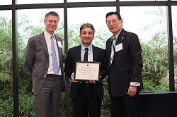
Amir H. Goldan named winner of the Stony Brook University Chapter of the National Academy of Inventors.
Dr. Goldan is an Assistant Professor in the Department of Radiology, and is an adjunct faculty member of the ECE department. He received his Ph.D. in Electrical and Computer Engineering at the University of Waterloo in 2011. Dr. Goldan has 5 issued and 2 pending patents in the field of medical imaging detectors and he received NAI award "For his innovative development and fabrication of medical imaging detectors for positron emission tomography (PET) and digital mammography.”
For more information, please click here.
Grant award
May 2018
A team of imaging scientists, radiologists, oncologists were awareded a $100,000 Mt Sinai-Stony Brook pilot grant to study breast lymph-node metastasis and response to therapy. Investigators: Duong, Cohen, Fisher, Palermo, Bernstein, Brian O'Hea (Stony Brook), and Shapiro, Margolies, Port, Schmidt (Mt Sinai)

National Distinguished Investigator Award to SBM Radiology Faculty
Apr 2, 2018
Dr. Tim Duong, PhD, Professor and Vice Chair for Research, is awarded with the 2018 Distinguished Investigator Award 2018 from The Academy for Radiology & Biomedical Imaging Research.
He wil also be inducted into the Academy’s Council of Distinguished Investigators at the 2018 Radiological Society of North Americas (RSNA).

Another Milestone in the Development of Computed Tomography Colonography (Virtual Colonoscopy)
February 2018
Collaboration between the research teams of Stony Brook University (led by Professor Jerome Z. Liang, PhD) and Wisconsin School of Medicine (led by Professor Perry J. Pickhardt, MD) has reached another milestone of developing computed tomography colonography (CTC), also known as Virtual Colonoscopy, to detect and understand the polyps as a precursor of the deadly colorectal cancer (CRC), which remains the 2nd leading cause of cancer occurrence and cancer-related death. This milestone is a NIH R01 award over $3M, entitled “Radiogenomics of Colorectal Polyps to Assess Benign Proliferative vs. Premalignant States” and led by Professor Pickhardt, to explore a conjecture that many gene mutations may occur very early in polyp formation rather than sequentially (i.e. the “Big Bang” hypothesis of tumorigenesis) and some polyps might be “born to be bad”.
Two examples of previous milestones include: (1) The recent NIH R01 award over $2M, entitled “Advancing Virtual Colonoscopy for Early Cancer Screening” and led by Professor Liang, to advance the CTC’s current paradigm of only detecting polyps toward also differentiating malignant polyps from non-malignant ones in a cost-effective fashion, whch can be read about ; and (2) The recommendation of CTC as a clinical option, by the U.S. Preventive Services Task Force, for screening of early colorectal cancer, which can be read about here.

Stony Brook Faculty Authored PET/MR Imaging Book
January 2018
Four of our Faculty members; Robert Matthews, MD, Lev Bangiyev, MD, Dinko Franceschi, MD, Mark Schweitzer, MD and one of our former residents; Rajesh Gupta, MD, have authored a book titled 'PET/MR Imaging' which was just published by Springer.
The book offers an overview of the clinical applications of PET/MR imaging through a case-based format. Each case in the book is presented with the patient history, protocols, interpretation of findings, and pearls and pitfalls accompanied by high quality PET/MR images.
To learn more about the book, please click here.
NIH Grant Funding
January 2018
Dr. Amirhossein Goldan is awarded his second NIH-R21 (NIBIB Trailblazer) grant for the feasibility study of a novel high-efficiency avalanche detector for high resolution x-ray imaging applications.
The proposed detector features a very thin layer of ultra-high efficiency Cadmium Selenide (CdSe), while coupled to a thicker multi-well avalanche amorphous selenium (a-Se) film, to serve as the photoelectric conversion layer for further enhancing the low-dose x-ray sensitivity (in addition to having avalanche gain inside a-Se). This project is inspired by the commercial success of solution-processed and highly tunable colloidal quantum dots (e.g., release of the first Quantum-Dot LED or QLED TV in 2015) for the formation of the CdSe layer. This project is in close collaboration with Professor Wei Zhao of Radiology who is also the Co-PI on this 3-year grant.
For more information, please click here.

