Lihong V. Wang, Ph.D., Bren Professor >Kris Krishna Kandarpa, M.D., Ph.D. >Bahaa Ghammraoui, PhD >Yvonne W. Lui, MD >

Abstract: We developed photoacoustic tomography (PAT) to peer deep into biological tissue. PAT provides in vivo functional, metabolic, molecular, and histologic imaging across the scales of organelles through organisms. We also developed light-speed compressed ultrafast photography (CUP) to record 219 trillion frames per second, orders of magnitude faster than commercially available camera technologies. CUP can record in real time the fastest phenomenon in nature, namely, light propagation for the first time and can be slowed down for slower phenomena such as neural conduction. We are exploring quantum entanglement for imaging.
PAT physically combines optical and ultrasonic waves. Conventional high-resolution optical imaging of scattering tissue is restricted to depths within the optical diffusion limit (~1 mm). PAT beats this limit and provides centimeter-scale deep penetration at high ultrasonic resolution and high optical contrast by sensing molecules. Broad applications include early-cancer detection and brain imaging. The annual conference on PAT has become the largest in SPIE’s 20,000-attendee Photonics West since 2010.
CUP with a single exposure can image transient events occurring on a time scale down to 10s of femtoseconds. Akin to traditional photography, CUP is receive-only—avoiding specialized active illumination required by other single-shot ultrafast imagers. CUP can be coupled with front optics ranging from microscopes to telescopes for widespread applications in both fundamental and applied sciences, ranging from biology to cosmophysics. Quantum imaging at the Heisenberg limit improves spatial resolution linearly with the number of quanta, faster than the square-root standard quantum limit.
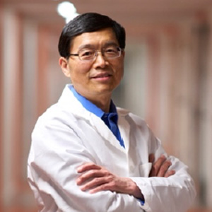
Biography: Lihong Wang earned his Ph.D. degree at Rice University, Houston, Texas under the tutelage of Robert Curl, Richard Smalley, and Frank Tittel. He is Bren Professor of Medical Engineering and Electrical Engineering, Andrew and Peggy Cherng Medical Engineering Leadership Chair, and Executive Officer (aka Department Chair) of Medical Engineering at California Institute of Technology. His book entitled “Biomedical Optics: Principles and Imaging,” one of the first textbooks in the field, won the 2010 Joseph W. Goodman Book Writing Award. He also edited the first book on photoacoustic tomography. He has published 590 peer-reviewed articles in journals, including Nature (Cover story), Science, PNAS, and PRL, and has delivered 580 keynote, plenary, or invited talks. His Google Scholar h-index and citations have reached 152 and 100K, respectively. His laboratory was the first to report in vivo/functional photoacoustic tomography (Top 2 cited in photoacoustics), 3D photoacoustic microscopy (Top 2 cited in photoacoustics), photoacoustic endoscopy, photoacoustic reporter gene imaging, the photoacoustic Doppler effect, the universal photoacoustic reconstruction algorithm (widely adopted), microwave-induced thermoacoustic tomography, ultrasound-modulated optical tomography, time-reversed ultrasonically encoded optical focusing, compressed ultrafast photography (70 trillion frames/s, world’s fastest real-time camera), and open-source Monte Carlo simulation of light transport in tissues (widely cited, celebrated by the Journal of Biomedical Optics in 2022). Photoacoustic imaging broke through the long-standing diffusion limit on the penetration of optical imaging, providing the only technology for noninvasive multiscale biochemical, functional, and molecular imaging from organelles to humans at high resolution. The technology has been commercialized by dozens of companies for both preclinical and clinical imaging (FDA approved for breast imaging). He chairs the annual conference on Photons plus Ultrasound, the largest conference at the annual 20,000-attendee Photonics West. He was the Editor-in-Chief of the Journal of Biomedical Optics. He received the NSF CAREER, NIH FIRST, NIH Director’s Pioneer, NIH Director’s Transformative Research, and NIH/NCI Outstanding Investigator awards. He also received the OSA C.E.K. Mees Medal, IEEE Technical Achievement Award, IEEE Biomedical Engineering Award, SPIE Britton Chance Biomedical Optics Award, IPPA Senior Prize, and OSA Michael S. Feld Biophotonics Award for “seminal contributions” to photoacoustic tomography and light-speed imaging. He is a Fellow of the AAAS, AIMBE, Electromagnetics Academy, IAMBE, IEEE, OSA, and SPIE as well as a Foreign Fellow of COS. An honorary doctorate was conferred on him by Lund University, Sweden. He was inducted into the National Academy of Inventors and the National Academy of Engineering.

Abstract: This talk will discuss trends & accomplishments in various medical imaging technologies funded by NIBIB. Sample projects will be highlighted to demonstrate innovative approaches in advancing medical imaging. Recent major developments in Artificial Intelligence applications in medical imaging will also be discussed.
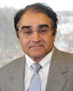
Biography: Kris Krishna Kandarpa, M.D., Ph.D. (Engineering Science.), is a cardiovascular and interventional radiologist and Director of Research Sciences and Strategic Directions at National Institute of Biomedical Imaging & Bioengineering /NIH. Prior to joining the NIH, he was CMO/CSO/EVP-R&D at Delcath Systems, Inc., Manhattan, NY, a manufacturer of minimally invasive combination device/drug treatments for liver tumors. Prior to his industry sojourn, he served as a tenured Professor & Chair of Radiology, University of Massachusetts Medical School & Radiologist-in-Chief of UMass-Memorial Health Care, where he established the Center for Innovative Imaging & Intervention. Earlier, at the Weill Medical College of Cornell University, he was a Professor of Radiology, Chief of Service & CVIR Director at The New York Presbyterian Hospital, and simultaneously an adjunct professor at the College of Physicians and Surgeons of Columbia University. Prior to New York, he was associate professor at the Harvard Medical School & co-director of CVIR at the Brigham & Women's Hospital, while also serving on faculty member at the Harvard-MIT Health Sciences & Technology Program, in Boston, MA. Dr. Kandarpa was an active biomedical researcher during his academic carrier, has published extensively, and lectured world-wide. He has served as Chair of the Research & Education Foundation of the Society of Interventional Radiology (SIRREF) and was on the board of the Academy of Radiology Research (ARBIR), when it helped establish NIBIB at the NIH. He is the author a text book entitled Peripheral Vascular interventions, which has been translated into Chinese and the popular Handbook of Interventional Radiologic Procedures, whose 6 th edition is to be released in May 2023, and which has been translated into 6 foreign languages.

Abstract: In this keynote speech, I will discuss the critical role of regulatory science in the field of medical imaging. I will focus on the work conducted at the FDA's Office of Science and Engineering Laboratories (OSEL) and Division of Imaging, Diagnostics, and Software Reliability (DIDSR). I will discuss the urgent need for new tools to assess AI algorithms in medical imaging, including the potential limitations of traditional testing methods in the age of AI, and the accuracy of CAD algorithms using novel deep learning techniques. I will touch upon the importance of pediatric-specific evaluations and the development of task-based assessments for deep learning in CT denoising. Additionally, I will highlight the advancements in using computational modeling for device evaluation, specifically through the VICTRE project. I will conclude by discussing the advancements in evaluating spectral x-ray imaging systems with photon-counting detectors. All of the above will be anchored in the current regulatory landscape for medical imaging devices and will include resources to relevant FDA Guidance documents.
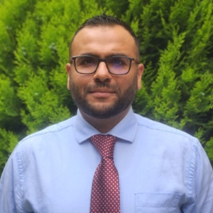
Biography: Dr. Ghammraoui is a Medical Imaging Scientist at the US Food and Drug Administration. He conducts research in medical imaging modalities and imaging physics to support FDA's regulatory mission. He has a particular focus on evaluating spectral imaging and AI applications in CT. He is also the acting liaison to IEC TC62 Working Group #44, where he actively participates in the development of standards for dual-energy X-ray radiography.
He earned his PhD in Physics from the National Institute of Applied Sciences in Lyon, France, in 2012. He completed his research at the Atomic Energy Commission's LETI Laboratory in Grenoble.
He is an active contributor to his field, having authored over 30 peer-reviewed articles on X-ray spectral imaging and holding 4 patents. He has also shared his insights at numerous national and international conferences, particularly regarding the intersection of spectral imaging and AI applications in CT.

Abstract: Deep learning has quickly revolutionized MR image reconstruction and gone quickly from an idea to clinical reality with all major equipment vendors and multiple start ups entering the space. The impact on image quality and imaging speed has been impressive. I will discuss some basics of deep learning-based MR reconstruction as well as some deep learning-based image enhancement as well as talk about the limitations and discuss some novel possible ways forward.
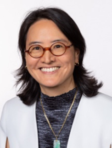
Biography: Yvonne W. Lui, MD is Professor and Vice Chair of Research for the Department of Radiology at NYU Grossman School of Medicine and the Vilcek Institute of Graduate Biomedical Sciences. She previously served as the Chief of Neuroradiology in the department for 7 years and as the inaugural Associate Chair for Artificial Intelligence, building an innovative program to leverage technological advances in machine learning for medical imaging. A native of New York City, Dr. Lui is a graduate of Swarthmore College and Yale University where she studied Physics and Medicine. She completed her residency and fellowship at NYU. She leads a world-renown radiology research program known for being a leader in imaging technology development and innovative translational research. The program ranks consistently in the top 10 in the nation for NIH-funding and there are over 35 faculty and 100 non-faculty personnel including postdoctoral fellows and research scientists. She serves as the collaboration lead on major research partnerships with both academia and industry including NYU Courant Institute and Center for Data Science, Siemens Healthineers, and Facebook AI Research. Dr. Lui is a NIH-funded researcher in translational neuroimaging to study traumatic brain injury. She serves on multiple NIH scientific review committees, is Past President of the New York Roentgen Society (NYRS), former oral board examiner for the American Board of Radiology (ABR) and a member of their Standard Setting Committee, member of the board of directors of the American Society of Neuroradiology (ASNR), and Senior Editor for the American Journal of Neuroradiology (AJNR). She is the current President of the ASNR.
