Located in a Level I Trauma Center, which designates the highest standard for surgical care, we work to explore and utilize computational and engineering tools to improve neuromodulation treatments for patient populations with severe traumatic brain injury (sTBI). Traumatic brain injury results in severe morbidities and mortality and accounts for more than 61,000 deaths every year(1). Coma and disorders of consciousness are common after TBI. A current lack of prognosis not only makes it difficult to counsel family members but hinders our ability to measure whether prospective treatments can be proven effective.
Our lab’s main focus is to develop a computational framework to understand the dynamical system that underlies different brain states (coma and consciousness states) and utilize stimulations to effectively improve the brain state from a pathological state to a healthy state. To do so, we utilize functional neurosurgery techniques in combination with imaging, computational neuroscience, and state-of-the-art machine learning tools. We work closely with our collaborators in Radiology, Electrical Engineering, Material Science, Applied Mathematics and Statistics, and Psychology to tackle this complex problem and accomplish this ambitious mission.

Detecting the Recovery of Consciousness After Brain Injury
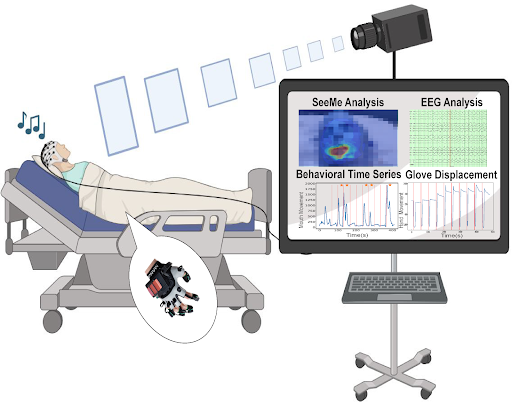
Our innovative project integrates cutting-edge computational neuroscience, computer vision, and neuromodulation to transform the way we diagnose, monitor, and treat disorders of consciousness. By developing “SeeMe,” a high-resolution system that leverages advanced computer vision algorithms and sensors, we can detect subtle, low-amplitude responses to auditory commands well before traditional clinical assessments. This approach is paired with advanced neural recordings (EEG, ECoG) and machine learning to uncover objective biomarkers that not only guide clinical decision-making but also serve as robust indicators for the effectiveness of emerging therapies.

Developing Non-Invasive Imaging Methods to Detect Brain AVM Before Rupture
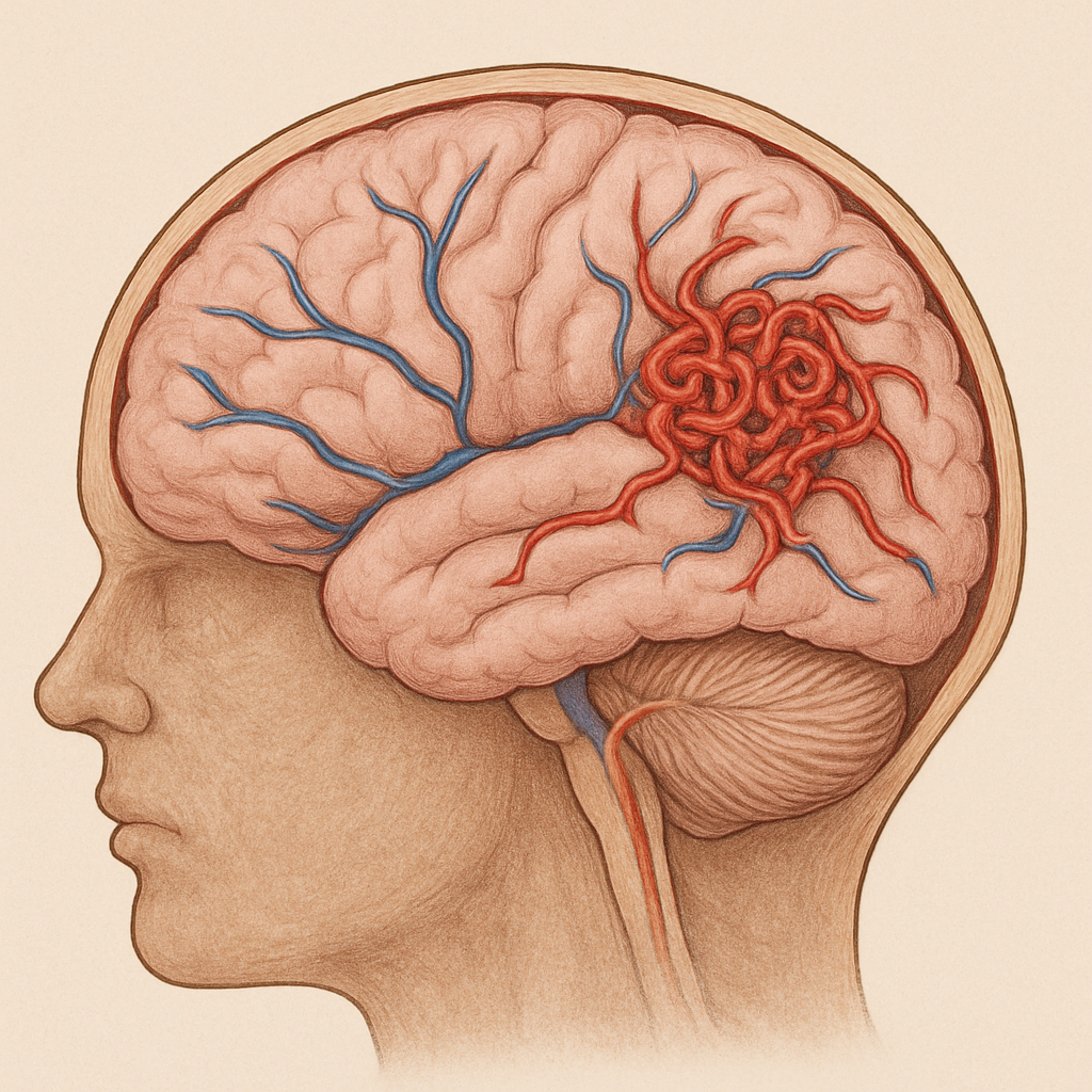
Our lab is advancing the diagnosis of brain arteriovenous malformations (AVMs) through cutting-edge research focused on non-invasive imaging and hemodynamic analysis. We focus on leveraging non-invasive imaging techniques, such as Doppler ultrasound, to study hemodynamic patterns and identify reliable diagnostic markers. Our research includes studies on in vitro models, 3D-printed modeling, healthy controls, and patients, exploring the utility of various diagnostic modalities in unruptured intracranial AVMs.
 Twitter
 Facebook