
 |
Professor Emeritus Director, Center for Infectious Diseases Ph.D., University of California-Berkeley, 1987 |
|||
|
Research |
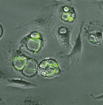
Dr. Bliska’s research seeks to understand molecular mechanisms that underlie pathogenesis or host protection during host-microbe cell interactions. In particular, he is interested in studying the activities of virulence factors that promote pathogenesis as well as trigger protective host innate and adaptive immune responses. The closely related Gram-negative bacteria Yersinia pseudotuberculosis and Yersinia pestis are used as model human pathogens to understand this dual nature of virulence factors. Y. pseudotuberculosis and Y. pestis are facultative intracellular pathogens and both use a highly conserved type III secretion system to deliver multiple effector proteins into the plasma membrane and cytoplasm of host cells. Numerous experimental approaches, including genetics, cell biology, immunology, structural biology, and functional evolutionary genomics, are used to determine how the type III secretion system and other Yersinia virulence factors work to promote disease or protective immunity. Specific questions being asked include: How do these bacteria prevent phagosome acidification and survive in macrophages? Which components of the type III secretion system are important for generating protective innate and adaptive immune responses in the host? How does the activity of one effector impact the immune response to another effector? Has allelic variation of virulence factors contributed to the process by which a bacterium with low pathogenic potential (Y. pseudotuberculosis) evolves into a bacterium with extremely high pathogenic potential (Y. pestis)?
|
|||