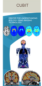
Under the direction of Christine DeLorenzo, PhD in Biomedical Engineering, the Center is a team of image analysts, programmers, and engineers, focused on extracting the most accurate quantitative information from medical images, with 10+ years of experience in the analysis of PET and MRI images including:
- External funding for PET and MRI imaging, including NIH and private foundations.
- Presentation of methodology and results at international conferences

- >100 imaging publications
- Development and validation of novel algorithms of image analysis
- Implementation of standard data analytical techniques
CUBIT facilitates image acquisition and analysis from Magnetic Resonance Imaging sequences including:
- structural MRI
- functional MRI (task and task-free)
- diffusion MRI (DTI and DSI)
- Arterial Spin Labeling (ASL, blood flow)
- Spectroscopy
We also aid in the acquisition and analysis of Positron Emission Tomography (PET) including:
- Drug Occupancy Studies
- Longitudinal Analysis
- Test-retest (stability) Analysis
In conjunction with the Bahl Radiochemistry, we will assist with imaging studies for any discipline (e.g. neurology, cardiology, oncology, etc.) or target (e.g. brain, heart, lung, liver, etc.) in humans, baboons or rodents.
Services
- Assistance preparing applications for internal pilot funding, government and private foundation groups
- Assistance developing imaging protocols
- Quality Assurance of PET and MRI images
- HIPAA compliant storage of demographic, clinical and image data that is backed up daily
- Raw image data files
- Transparent image analysis pipeline with web-based interface for viewing results
- Write-up of methodology for manuscripts
- Full PET quantification including
- Fitting of plasma and metabolite data
- Time activity curve generation
- Kinetic/Graphical modeling
- Regional and voxel analysis on a group level
Unique Equipment
Unique Stony Brook equipment, developed by Drs. Paul Vaska and Daniela Schultz
Rat Conscious Animal Scanner (RatCAP)
- allows for PET imaging of the rat brain while the rat is conscious and behaving
- field of view covering the whole rat brain (38 mm transaxial, 18 mm axial)
- spatial resolution of 1.5 mm FWHM
MR-Compatable RatCAP
An MR-compatible version of the RatCAP has been developed that can be installed within SBU's Bruker 9.4 T small-animal scanner
- custom-built head holder and bed with capabilities for gas anesthesia and physiological monitoring
- The PET system has similar performance as the RatCAP but also has inputs for respiratory and cardiac gating as necessary