THE MENINGES
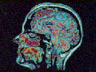
The meninges consist of three layers. The outermost is called the dura mater, also known as the pachymeninx (pachy = thick). It consists of two fused layers of dense fibrous connective tissue. The external layer is adherent to the skull and actually forms the periosteum. In older individuals, the dura can fuse with the skull.
In several locations, the two layers separate and form the dural sinuses, the major venous channels of the brain. The dura is folded in the midline to form the falx cerebri, which incompletely separates the two cerebral hemispheres. The other major fold is the tentorium cerebelli, which separates the cerebellum from the cerebral hemispheres. The tentorium stretches over the top of the posterior cranial fossa. When considering the location of pathological processes, it is common practice to separate them into supratentorial and infratentorial processes.
The potential space located between the dura and the bones of the cranium is known as the epidural space. The potential space between the dura and the underlying meninges and brain is called the subdural space. In life, there are no actual spaces, but these are tissue planes that may be separated.
The arachnoid and the pia mater together form the leptomeninges (lepto = slender). The arachnoid, a thin membrane, is connected to the underlying pia via wispy connective tissue fibers that resemble spider webs; these give the arachnoid ("spider-like") its name. The arachnoid is loosely applied to the brain and is normally almost transparent.
The pia is a delicate membrane that closely covers the brain surface, following all of its convolutions and coating the penetrating arterioles and venules to the point where they become capillaries. In the spinal canal, the pia forms the denticulate ligaments and the filum terminale. The pia is closely related to the peripheral layer of astrocytic end feet. Together, they form the pia-glial limiting membrane of the central nervous system. The pia is intimately connected to the surface of the brain and spinal cord, but some pathological processes, for example, certain brain tumors, can spread within and along the subpial space.
The space between the pia and arachnoid is the subarachnoid space, which is filled with cerebrospinal fluid (CSF). The brain and spinal cord are thus suspended from the dura-arachnoid by the arachnoid trabeculae and are floating in a bath of CSF. The subarachnoid space also contains the superficial cerebral arteries and veins. In areas where the brain and spinal cord surface is relatively far from the dura-arachnoid, the subarachnoid space is enlarged to form cisterns. The largest subarachnoid cistern surrounds the medulla and the base of the cerebellum. This is the cerebello-medullary cistern, or the cisterna magna. |
THE BRAIN
The brain has five major anatomic divisions that are based upon the structure of the developing brain. These are:
1. the telencephalon, which includes the cerebral hemispheres and the deep structures known collectively as the basal ganglia
2. the diencephalon, which is composed predominantly of the thalamus, epithalamus and hypothalamus
3. the mesencephalon, or midbrain
4. the metencephalon, consisting of pons and cerebellum
5. the myelencephalon, which is the medulla |
EXTERNAL FEATURES OF THE BRAIN
When looking at the brain, the cerebral hemispheres are the most prominent structures of the brain. The cerebral cortex, which is located on the surface is highly convoluted. This allows for the packing of a great surface area in a small space (the cranial cavity). In other animals, such as rats, rabbits and frogs, there has not been such a great expansion of the cortex and these brains are smooth (lissencephalic). The elevated folds are called gyri, while the furrows between them are called sulci. Very deep sulci are also referred to as fissures. While there is some inter individual variability in the patterns of gyration and sulcation, there is actually a striking consistency in the pattern.
| Using the images of the lateral and superior views of the cerebral hemispheres, you should review the locations of the Sylvian (lateral) fissure and the central sulcus . The precentral gyrus (with primary motor cortex) and the postcentral gyrus (with primary somatosensory cortex) are anterior and posterior to the central sulcus, repectively. The important language areas, Broca's and Wernicke's areas in the dominant hemisphere. Appreciate the normal appearance of gyri and sulci. |
|
| On the inferior view of the brain, identify the optic chiasm and mammillary bodies. Other than their distal stumps, the optic nerves are not present in the specimen. Identify the pons, medulla and cerebellum. |
 |
|
Internal neuroanatomical landmarks and highlights of normal histology of brain
| In the coronal sections 1, 2, & 3 review the locations of the basal ganglia, which include the caudate nucleus, the putamen and the globus pallidus. Identify the thalamus, amygdala and hippocampus. Examine the appearance of the subcortical white matter, corpus callosum and internal capsule. Note the locations, size and shape of the lateral and third ventricles. The lateral ventricles in this brain are mildly dilated. |
|
The parenchymal cells of the central nervous system are neurons and glia. The important cell types to examine are neurons, particularly cerebral cortical pyramidal neurons, oligodendroglia, astrocytes, ependymal cells and choroid plexus epithelium. The latter four cell types are glia or derived from glia. One should also examine leptomeninges and blood vessels.
| View a histological section of Cerebral cortex with subcortical white matter. This is stained with Hematoxylin and Eosin (H&E), a routine, general purpose histological stain used for basic examination of tissue. Note that the cortex is a slightly paler pink than the more deeply staining subcortical white matter. |
 |
| First view the normal arachnoid and the subarachnoid space that contains normal superficial blood vessels that supply the brain. Identify the cerebral cortex that contains six layers of neurons. Layer 1 is the most superficial and layer 6 is deepest. Layers III and V are richest in pyramidal neurons. Note the normal texture of the neuropil between the neurons. Examine the cerebral cortex at higher power and identify pyramidal neurons with their large nuclei and prominent nucleoli. Notice the basophilic (blue) character of the cytoplasm of these cells, which is a characteristic feature of neurons. Many of the pyramidal neurons have small round nuclei associated with them. These round nuclei are known as satellite cells, which are oligodendroglial cells in gray matter. |
|
| The subcortical white matter is largely composed of axons and oligodendroglia. In the white matter, the oligodendroglia are the myelin-forming cells of the central nervous system. Note their characteristic round nuclei, surrounded in many cells by a clear rim. This characteristic "fried-egg" appearance of oligodendroglial cells is actually an artifact of delayed fixation. It is an artifact that is very useful for the identification of these cells. Notice the cellularity and texture of normal white matter. Look at normal blood vessels. |
|
| Look at a section of basal ganglia at low and higher magnifications. The caudate and putamen are histologically identical because of their common developmental origin. The caudate is easy to identify because it is located in the lateral wall of the lateral ventricle, so it will always have an ependymal lining. Examine the ependyma, which consists of a single layer of ciliated cuboidal epithelium at the ventricular surface of the brain. It also lines the central canal of the spinal cord. The central canal is normally not an open structure in adults as it is in the developing central nervous system. Examine the caudate and putamen . The globus pallidus has a different texture and different cellular composition than the caudate and putamen, which goes along with its differing neurophysiological function. |
|
| The hippocampal formation has three components: the dentate gyrus, the hippocampus proper (also called Ammon's horn) and the subicular complex. The dentate gyrus contains a layer of small neurons. Ammon's horn has four sections, CA1-4. The CA1 region is important to recognize because of the particular vulnerability of the neurons in the pyramidal layer to global hypoxic-ischemic events. |
|
| The midbrain, or mesencephalon, is the location of the cerebral aqueduct (of Sylvius), the corpora quadrigemina (superior and inferior colliculi), the substantia nigra and the cerebral peduncles. Note the normal appearance of the pigmented neurons of the substantia nigra. The oculomotor nuclei are located here and the oculomotor nerves course ventrally to exit at the interpeduncular fossa. The trochlear nuclei and nerves are located in caudal midbrain. |
 |
 |
| The pons has a tegmentum and base. The locus ceruleus is an important pigmented noradrenergic nucleus located lateral to the fourth ventricle in the tegmentum. The base contains the crossing pontocerebellar fibers that form the middle cerebellar peduncle and the corticospinal tracts run through the base of the pons on their way to the spinal cord. The cranial nerves and nuclei of the pons are V- VIII. |
|
| A section of spinal cord reveals the dorsal and ventral roots, the dorsal horn, the ventral horn which contains the spinal motor neurons and the white matter tracts located at the periphery of the cord. Note the typical appearance of a motor neuron, with its prominent nucleolus in the nucleus and the basophilic (blue in H&E stain) tigroid appearance of its cytoplasm, which is due to the presence of abundant ribosomes needed in the manufacture of proteins |
|
|
|

