The following are images of representative pathologic conditions of the genital tract. They have been made from histologic slides that are present in the Reproductive System Laboratory slide boxes that are distributed. The examples represent a sampling and are not all inclusive of diseases that will be discussed or that you are expected to know. The laboratory syllabus provides a more detailed discussion of each of the cases.
Click on each image for larger version.
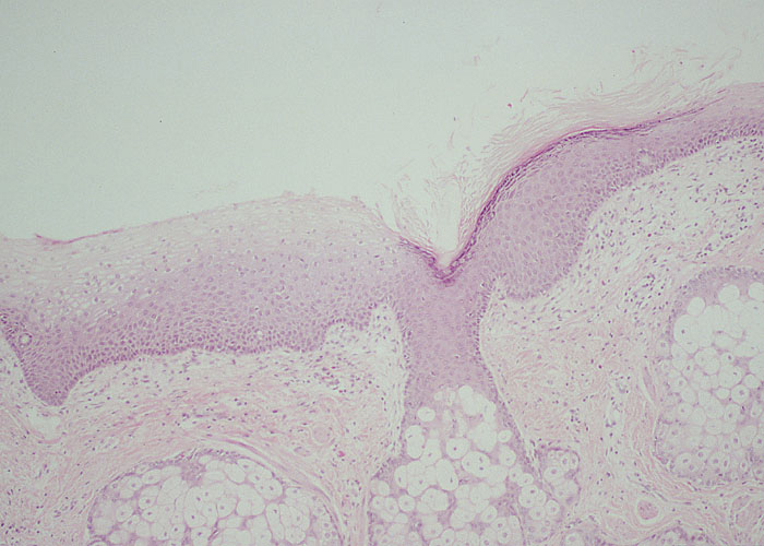
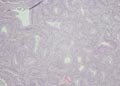
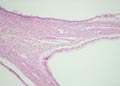
Cervical Dysplasia
(Slide 14)
| 
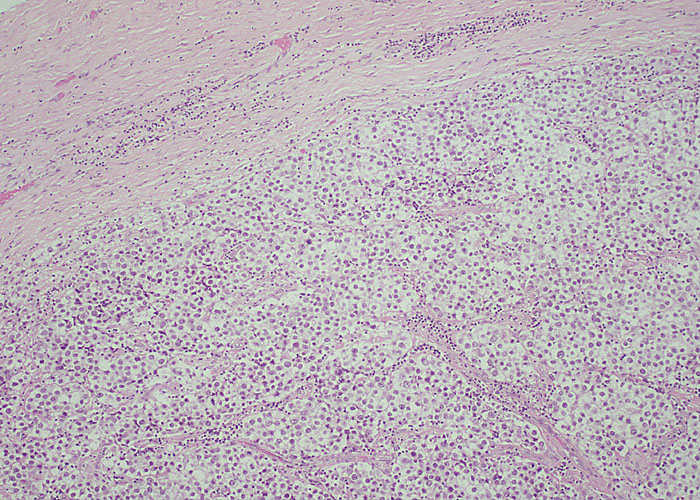
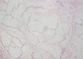
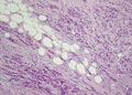
Dermoid Cyst of the Ovary
(Slide 66)
|

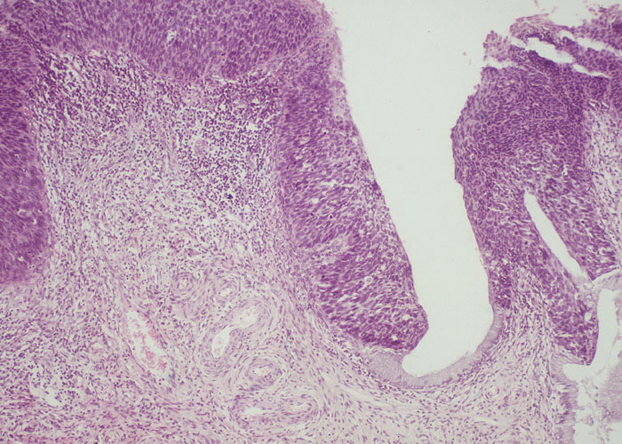
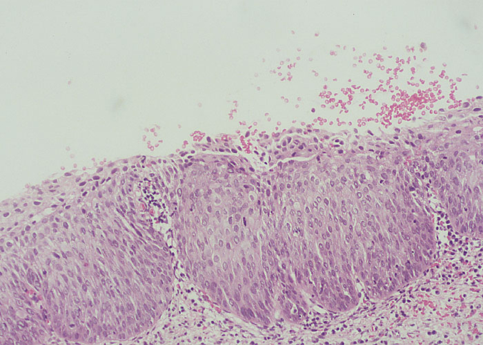
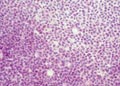
Cervical Carcinoma In-Situ
(Slide 15)
| 
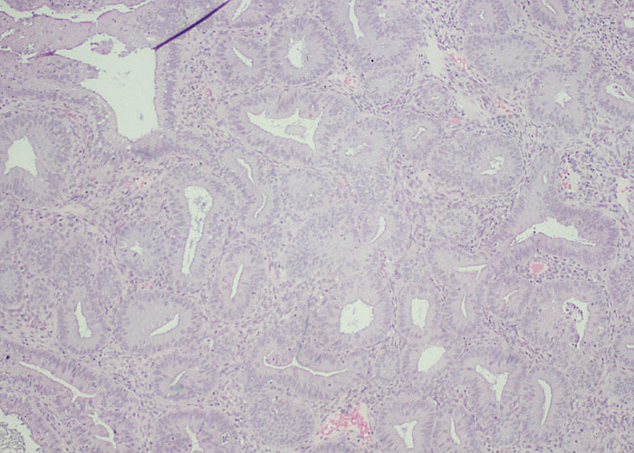
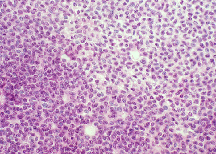
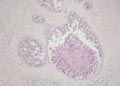
Dysgerminoma of the Ovary
(Slide 67)
|
Endometrial Hyperplasia
(Slide 34)
| 
Fibroadenoma of the Breast
(Slide 90)
|
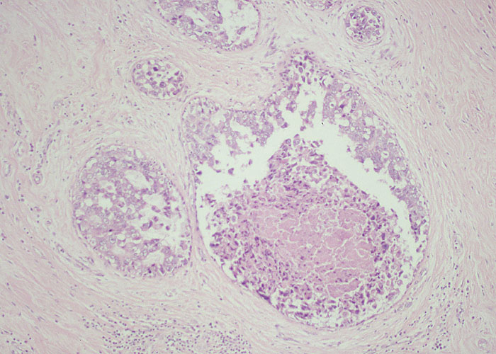
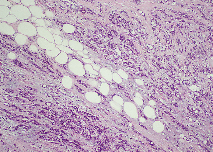
Granulosa Cell Tumor of the Ovary
(Slide 55)
| 
Intraductal Carcinoma of the Breast (Slide 92)
|
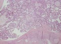
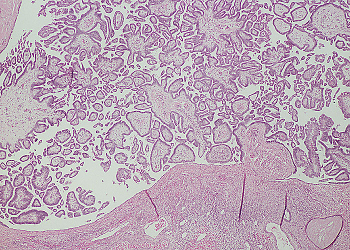
Ovarian Serous Borderline Tumor
(Slide 59)
| 
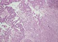
Invasive Mammary Carcinoma
(Slide 93)
|
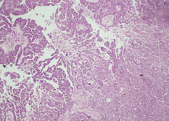
Serous Cystadenocarcinoma of the Ovary (Slide 61)
| 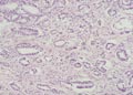
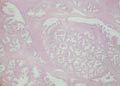
Adenocarcinoma of the Prostate
(Slide 96)
|
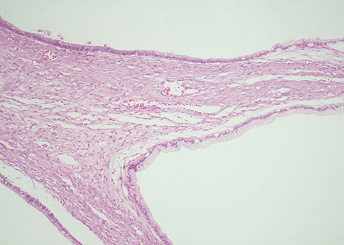
Mucinous Cystadenoma of the Ovary
(Slide 62)
| 
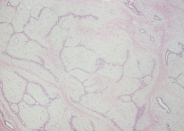
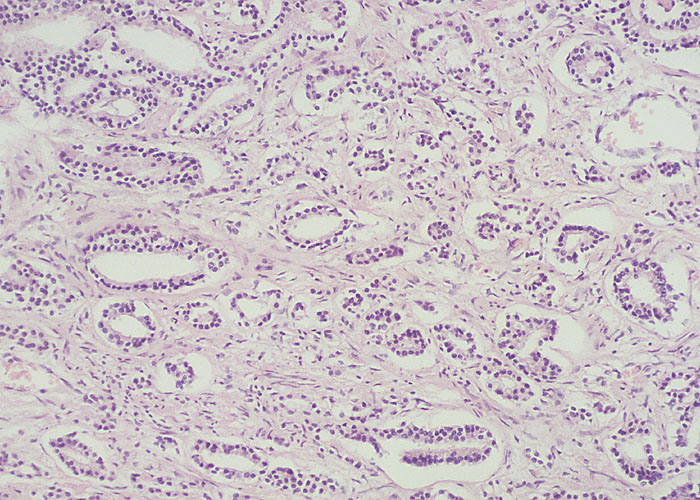
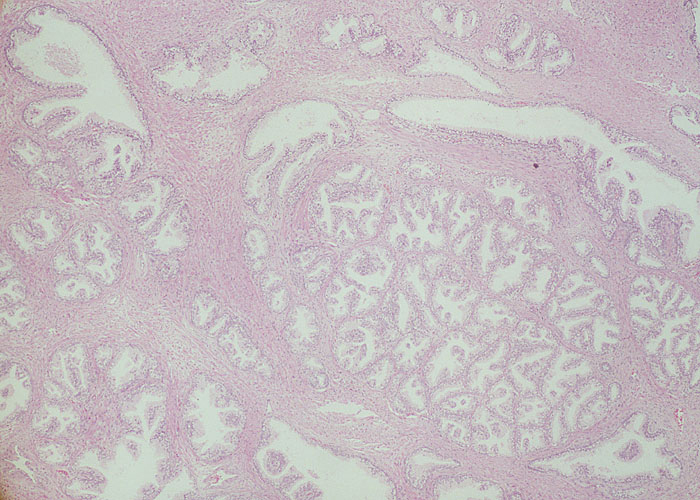
Benign Prostate Hyperplasia
(Slide 97)
|