Research
Stroke
Stroke is a leading cause of death and chronic disability worldwide. The available treatments for ischemic stroke remain limited. Recombinant tissue plasminogen activator (rt-PA) is effective but benefits only 3-5% of patients due to the risk of fatal hemorrhagic transformation and limited treatment window. Mechanical clot extraction has recently shown to be efficacious in clinical trials. There are currently no clinically approved neuroprotective drugs for treatment of acute stroke.
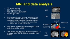 MRI offers a non-invasive means to track the progression of ischemic brain injury in a longitudinal fashion. T2-weighted MRI is widely used to visualize edema and define final infarct volume. Perfusion-weighted MRI can measure cerebral blood flow (CBF) at the tissue level, allowing for the detection of tissue with reduced perfusion that is at risk of ischemic brain injury. Diffusion-tensor imaging (DTI), which measures water motion, is very sensitive to early ischemic brain injury in contrast to computed tomography and T2 MRI. As such DTI has become the method of choice for early detection of ischemic brain injury. The combined use of perfusion and diffusion MRI are now widely used to distinguish reversible from irreversible ischemic brain injury, and to guide acute stroke treatment in preclinical and clinical settings.
Studies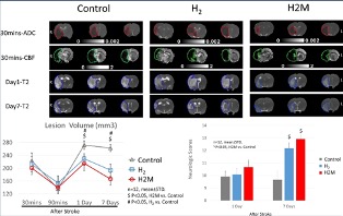
1. Hydrogen or minocycline individually have been shown to be neuroprotective in experimental ischemic stroke. This study evaluated the efficacy of hydrogen treatment in a rat stroke model compared to sham-treated controls using multiparametric MRI and neurological tests.
Additionally, comparison of H2 treatment alone was made with H2 combined with minocycline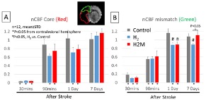 (H2M) treatment. The primary findings were: i) H2 therapy reduced infarct volume in both H2 and H2M groups compared to controls at 1 and 7 days after stroke, and ii) both H2 and H2M improved neurologic functional recovery on day 7. The secondary outcomes were: iii) H2M treatment attenuated post-stroke hyperperfusion in hyperacute phase, and iv) H2 minimized and H2M markedly minimized white matter injury. H2M outperformed H2 alone.
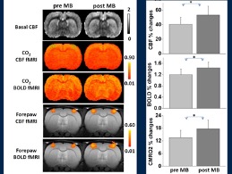
2. Methylene blue (MB) at low doses has metabolic-enhancing and antioxidant properties and exhibits experimental neurotherapeutic benefits. This study used MRI to evaluate the in vivo effects of a single intravenous MB therapeutic dose on basal CBF, blood oxygenation level-dependent (BOLD) and CBF responses to hypercapnic (5% CO2 in air) inhalation, as well as changes in BOLD, CBF, and cerebral metabolic rate of oxygen consumption (CMRO2) during forepaw stimulation in the rat brain. We found that this MB therapeutic dose did not have significant effects on arterial oxygen saturation, heart rate and fMRI responses to hypercapnia. However, MB significantly potentiated forepaw-evoked BOLD and CBF changes under normoxia. To further evaluate in vivo effects of MB under metabolic stress conditions, MRI measurements were also made under mild hypoxia (15% O2). Hypoxia increased evoked functional MRI responses. MB under hypoxia further potentiated forepaw-evoked BOLD, CBF and oxygen consumption responses relative to normoxia.
Cancer
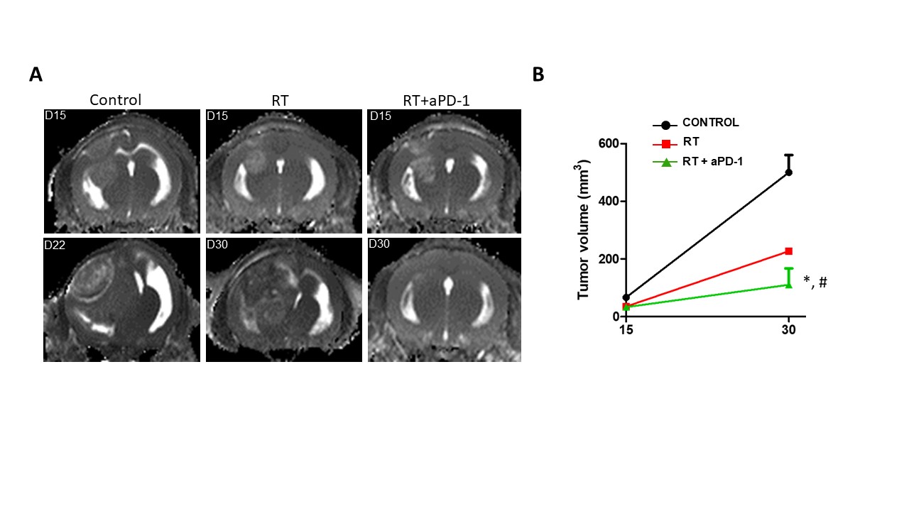 1. Glioblastoma multiforme (GBM) carries a grave prognosis despite aggressive treatment. We used multimodal MRI to longitudinally monitor changes in brain tumor volume growth due to the effects of a combination treatment of radiation therapy (RT) and anticancer drug in a mouse model of GBM. We showed that the combination therapy was more effective in reducing tumor burden and yielded better survival compared to non-treated or radiation-only.
(This project is collaborating with Dr. Samuel Ryu from the Department of Radiation Oncology at Stony Brook University Hospital)
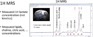
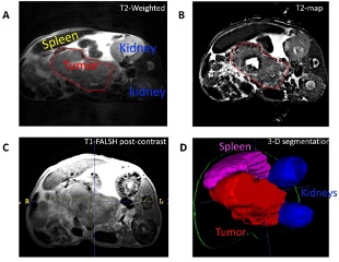 2. The lack of effective treatment options and intrinsic chemoresistance to widely used first-line therapeutic agents underlie the low survival of patients with pancreatic ductal adenocarcinoma (PDAC). Using Magnetic Resonance Imaging and Spectroscopy (MRI/MRS) in different subtypes of a PDAC animal model, we detected differences in tumor volume and in several metabolites, including the glycolytic activity indicator, lactate, suggesting that this imaging method provides information about pancreatic tumor metabolism in vivo.
(This project is a collaboration with Dr. Kenneth Shroyer from the Department of Pathology at Stony Brook Medicine and Dr. Luisa Escobar-Hoyos from the Department of Therapeutic Radiology, Yale Medicine)
Traumatic brain injury
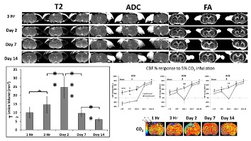 In traumatic brain injury (TBI), the initial physical impact causes direct mechanical damage, followed by progressive secondary injury due to brain swelling, cerebral blood flow disruption and metabolic dysfunction. In a rat model of TBI, CBF, carbon-dioxide reactivity, diffusion and T2 MRI were acquired 1-3 hours, 2, 7, and 14 days following a controlled cortical impact to the unilateral somatosensory/motor cortex. Diffusion and T2 MRI were useful markers for tissue damage and lesion detection.
New drug exploration
BOLD functional MRI can be used to study the neurovascular response to experimental drugs. Decreases in the BOLD signal of the whole brain were detected following injection of a new drug in mice. This approach will allow evaluation of activation/inhibition patterns of experimental drugs.