Top Five Mos t Cited Papers for Jerome Liang, PhD
December 20, 2016
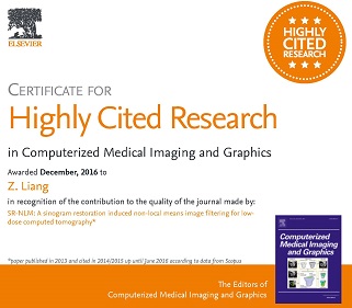
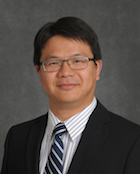
One research paper of Jerome Liang’s Lab, published in 2013 in the journal of Computerized Medical Imaging and Graphics, has been rated as the top 5 most cited papers from 2014, 2015 and 2016.
MSK Fellow to Receive 'Invest in Youth' Award
December 2016
Our MSK Fellow, Corey Ho, MD, has just received the award 'Invest in Youth' from the European Society of Radiology. Out of 3,000 Radiologists in training who applied for the award, our very own Dr. Ho, was chosen to receive it. He will receive the award at the ECR Conference in March 2017, where he will also be giving two oral presentations.
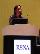
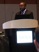
Physician Presentation at RSNA
November 30, 2016
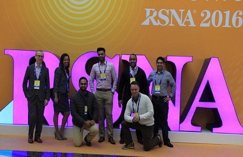
Drs. Robert Matthews, Lev Bangiyev and Dinko Francheschi presented at the Annual RSNA Conference in Chicago, IL this year.
“Incidental Brain Pathology on Whole Body FDG PET-MRI.” Franceschi AM, Matthews R, Bangiyev L, Chaudhry A, Relan N, and Franceschi D. Read more of their research.
Our Residents Presentations from the 2016 Annual RSNA Conference
November 27-30, 2016
Jerrin Varghese, MD
“CRMO Revisited: Do MRI Features Correlate with Clinical Response?”
Jerrin Varghese, MD | Marco A. Oriundo Verastegui, MD | Julie Cherian | Mingqian Huang, MD
- Dr. Varghese was also a recipient of the RSNA Travel Award
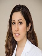
 Elham Safaie, MD
“Added Value of SPECT-CT to Standard Dynamic Imaging in Abdominal Emergencies”
Elham Safaie, MD | Kavitha Yaddanapudi, MD, DMRD | George C. Angelos, MD | Robert Matthews, MD
Ian Whiteside, MD
“Development and Evaluation of Natural Language Processing Software to Produce a Summary of Inpatient Radiographic Findings Identified for Follow-Up” 
Ian R. Whiteside, MD | Iv Ramakrishnan, PhD | Ritwik Banerjee, PhD | Vasudev Balasubramanian | Basava Raju Kanaparthi | Matthew A. Barish, MD
- Dr. Whiteside was also a recipient of the RSNA Travel Award
 Dharmesh Tank, MD
“Achilles Tendon Diffusion Tensor Imaging and Tendon Fiber Tracking by Stimulated Echo Resolve (ste-RESOLVE)”
Xiang He, PhD | Kenneth T. Wengler, MS | Chien-Hung Lin, MD | Marco A. Oriundo Verastegui, MD | Alex Sacher, BSC | Kevin S. Baker, MD | Mingqian Huang, MD | Elaine S. Gould, MD | Mark E. Schweitzer, MD | Dharmesh Tank, MD
 Jared Nesbitt, MD
“Value of Repeat Coronary CT Angiography in Patients Presenting with Acute Chest Pain”
Jared Nesbitt, MD | Christopher Shackles, DO, MPH | Jie Yang | Eric J. Feldmann, MD | Donglei Yin, PhD | Kavitha Yaddanapudi, DMRD, MBBS
“Spontaneous Osteonecrosis of the Knee (SONK): The Role of MR Imaging in Predicting Clinical Outcome”
Jared Nesbitt, MD | Dharmesh Tank, MD | Marco A. Oriundo Verastegui, MD | Elaine S. Gould, MD | Mingqian Huang, MD
- Dr. Nesbitt was also a recipient of the RSNA Travel Award
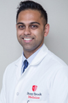
 Anuj K. Rajput, MD
“Medical Student Perception of Diagnostic Radiology after Implementation of an Evening Emergency Radiology Rotation”
Anuj K. Rajput, MD | Don N. Nguyen, MD | Toshie Ahluwalia, MD | Michael Goodman, BA | Rayeed Islam, BS | Jared Nesbitt, MD | Dharmesh Tank, MD | Robert Matthews, MD
- Dr. Rajput was also a recipient of the RSNA Travel Award
 Krystal D. Airola, MD
“Prevalence of Coronary Artery Disease (CAD) in Premenopausal Women Presenting with Acute Chest Pain Diagnosed by Coronary CT Angiography”
Krystal D. Airola, MD | Jie Yang | Donglei Yin, PhD | Kavitha Yaddanapudi, DMRD, MBBS | Dharmesh Tank, MD
- Dr. Airola was also a recipient of the RSNA Travel Award
Our 2016 MSK Fellowship Graduate also presented this year
 Matthew Teng, MD
"MSK Radiologist and the Orthopedic Surgeon Tag Team: A survey of MSK imaging training amongst Orthopedic Surgeons"
Teng, M | Nesbitt, J | Wang, K | Gould, E | Tank, D | Penna, J | Schweitzer, M
Medical Image Computing: We are living in interesting times
December 6, 2016
 The departments of Computer Science and Biomedical Informatics, along with Radiology's Dr. Mary Saltz, hosted a presentation by Dr. Ron Kikinis. Dr. Kikinis is the founding Director of the Surgical Planning Laboratory, Department of Radiology, Brigham and Women's Hospital, Harvard Medical School, Boston, MA, and a Professor of Radiology at Harvard Medical School. The importance of Medical Image Computing is increasing rapidly. Image Informatics, Machine Learning, Radiomics and quantitative imaging are all contributing to the portfolio of tools and technologies being deployed to address the new challenges, which Dr. Kikinis spoke about in his presentation.
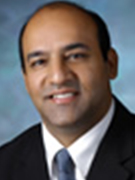
Dr. Shahid Hussain to be Named as a Fellow of the ACR
November 2016
 One of the highest honors the ACR can bestow on a radiologist, is recognition as a fellow of the American College of Radiology. ACR Fellows demonstrate a history of service to the College, organized radiology, teaching, or research. We would like to congratulate our own Dr. Shahid Hussain, on his honor of becoming a fellow of the ACR.
SPECT/CT can be Valuable in Abdominal Emergencies
November 10, 2016
 Radiology Resident, Elham Safaie, MD, has had a paper published on Aunt Minnie in regard to finding that when faced with a gastrointestinal or genitourinary imaging emergencies, adding SPECT/CT to the standard imaging options can help identify the source of
 the problem and lead to an appropriate intervention.
To learn more about this study, please click here.
Grand Rounds: "The Story of MRI, Its Technology and Its Most Recent Frontier in the CSF Physiology of the Brain"
November 10, 2016
 Dr. Raymond V. Damadian presented Grand Rounds on November 10, 2016, here in the Department of Radiology. His presentation was titled "The Story of MRI, Its Technology and Its Most Recent Frontier in the CSF Physiology of the Brain." Dr. Damadian is Professor of Medicine and Radiology at SUNY Downstate and is President, Chair and Founder of FONAR Corporation- a manufacturer of MR Scanning Systems. His research interests lie in MRI scanner technologies and clinical applications of MR Scanning.
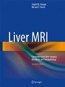
Liver MRI - Correlation with Other Imaging Modalities and Histopathology New Book Published by Dr. Hussain
November 2016
Dr. Hussain is an American Board of Radiology certified radiologist with clinical experience of more than 15 years and active in clinical research and education with 74 peer-reviewed papers, 44 abstracts, 162 presentations, and 2 MRI textbooks. The second edition of his textbook entitled “Liver MRI” was recently published in December 2015. This book provides a practical approach to liver MRI, with coverage of the most up-to-date MR imaging sequences, normal and variant anatomy, and diverse pathologic conditions. It features computer-generated drawings relating clinical concepts to the MRI findings, 2D and 3D reconstructions, relevant and systematic (differential) diagnostic information, numerous additional cases, recent literature references, and descriptions of patient management options. MRI findings are correlated to ultrasound, computed tomography, nuclear medicine exams, laboratory findings, and histopathology when appropriate.
To learn more, please click here.
Grand Rounds: "Cardiac MRI: Delayed Enhancement Patters and Beyond"
October 28, 2016
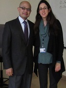 Dr. Alan C. Legasto, pictured here with Dr. Masha Hoshmand Kochi, presented Grand Rounds to our faculty and staff titled "Cardiac MRI: Delayed Enhancement Patters and Beyond". Dr. Legasto is a board-certified radiologist specializing in Thoracic Imaging. He is an Assistant Professor of Radiology at Weill Cornell Medical College and Assistant Attending Radiologist at the New York-Presbyterian Hospital-Weill Cornell Campus. In addition to clinical activities, Dr. Legasto is an active teacher of residents, fellows, and medical students. At Beth Israel Medical Center, Dr. Legasto was voted by the radiology residents as “Teacher of the Year” for three out of his eight years at Beth Israel. In 2013, he was selected as one of the “SuperDocs Rising,” a peer-elected acknowledgement for the New York Times Magazine.
Special Lecture: "The Invention and Applications of Avalanche Selenium Image Pickup Tube"
October 26, 2016
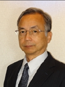 Kenkichi Tanioka, PhD presented a special lecture here in the Department of Radiology to our faculty and staff titled "The Invention and Applications of Avalanche Selenium Image Pickup Tube." Dr. Tanioka is a Visiting Professor here at Stony Brook in the Department of Radiology. He worked for NHK Science & Technology Research Laboratories (STRL), Tokyo, where he was engaged in the research of an amorphous selenium photoconductor for image pickup devices. He has contributed not only to the advanced broadcasting technologies but also to the fields other than broadcasting, e.g., observation of deep sea, living cells, X-ray images, etc. Dr. Tanioka is an inventor of the highly sensitive HARP pickup tube. He was awarded fifteen prizes including the Imperial Invention Prize in 1996.
Conference Presentation by Dr. Yaddanapudi
October 18, 2016
Dr. Kavitha Yaddanapudi presented 'Role of Coronary CTA in Evaluation of Premenopausal Female Patients Presenting to Emergency Department With Acute Chest Pain' at the 2016 Annual North American Society for Cardiovascular Imaging Conference.
Conference Presentation by Dr. Yaddanapudi
October 18, 2016
 Dr. Kavitha Yaddanapudi presented 'Role of Coronary CTA in Evaluation of Premenopausal Female Patients Presenting to Emergency Department With Acute Chest Pain' at the 2016 Annual North American Society for Cardiovascular Imaging Conference.
Conference Presentation by Dr. Feldmann
October 17, 2016
 Dr. Eric Feldmann presented 'The Triple-Rule-Out Study: Often Ordered, But Rarely Useful' at the 2016 Annual North American Society for Cardiovascular Imaging Conference.
New Faculty to Join the Department of Radiology
October 10, 2016
 Timothy Q. Duong, PhD, has joined the department as a Professor of Radiology and as the Vice Chair of Research. He received a BS in Biochemistry and (Physical) Chemistry from Stony Brook University. Then he went on to get his MS in Chemistry and his PhD in MRI and Diffusion MRI from Washington University. His Post Doctoral Training was in fMRI and Perfusion, which he did at the University of Minnesota. He has countless honors and awards from throughout the years and has mentored many PhD students. His research focuses on the development and application of magnetic resonance imaging (MRI), spectroscopy (MRS), and speckle and optical imaging, to the study of brain and retinal anatomy, physiology and function in animal models and humans. We are very excited to welcome him to the Department.
Conference Presentation by Dr. Fisher
October 1, 2016
 Dr. Fisher Co-Directed the 28th LIRS Breast Imaging Update Conference on October 1, 2016, in Melville, New York. He shared the podium with the Chief of Breast Imaging at the University of Pennsylvania, Dr. Emily Conant. Dr. Fisher presented on the topics of “Clinical Uses of Physics in Mammography,” “Contrast Mammography vs. MRI,” “Ongoing Controversy about Ultrasound Screening,” and presented interactive teaching files. This conference, sponsored by the LIRS, received 7.5 AMA PRA Category 1 CME Credits.
Dr. Barish's Publication to be Nominated for Presentation
October 2016
 "Visual Analytics of Image-derived Temporal Features — Focusing on the Spleen": A paper in which Dr. Matthew Barish is named in, has been nominated for presentation at in the TVCG Track at IEEE VAST in October 2016.
Mary's Musings: We Are All Imagers
September 30, 2016
 What does medical imaging have to do with deep see exploration? If your answer is "not much," then be sure to read this new column from Dr. Mary Saltz, who has just returned from a fascinating conference on new imaging technologies that span scientific disciplines. Click here to read more. (Log in required)
Dr. Baker named Residency Program Director
September 19, 2016
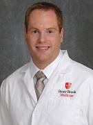 Stony Brook Department of Radiology is proud to announce that Kevin Baker, MD, has been named Residency Program Director for the Department of Radiology Residency Program. Dr. Baker graduated from Stony Brook University Summa Cum Laude with a Bachelor of Science in Biology, and Medical Doctorate. After completing his Internal Medicine Internship at Winthrop University Hospital, he completed his Residency, as Chief Resident, and his Musculoskeletal Fellowship at Stony Brook Medicine. The Graduate Medical Education Committee approved his appointment on Monday, September 19, 2016. The residents would like to congratulate Dr. Baker on this appointment, and are looking forward to his many years of program leadership.
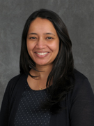
Grand Rounds: Cardiac MRI in Clinical Practice: Current Progress and Future Directions
September 29, 2016
 Monvadi Barbara Srichai-Parsia, MD, pictured here with Dr. Varghese Cherian, presented Grand Rounds to our faculty and staff titled 'Cardiac MRI in Clinical Practice: Current Progress and Future Directions'. Dr. Srichai-Parsia is an Associate Professor of Medicine and Radiology; Vice-Chief, MedStar Heart & Vascular Institute; Director, Cardiac Non-Invasive Imaging Laboratory at Georgetown University Medical Center.
Grand Rounds: "The Never Ending Controversy Surrounding Breast Cancer Screening"
September 2016
Dr. Fisher has been invited to give a grand rounds presentation to the Department of Family Medicine at Stony Brook Medicine, in September 2016. His topic is “The Never-Ending Controversy Surrounding Breast Cancer Screening.”
Grand Rounds: Cardiac MRI in Clinical Practice: Current Progress and Future Directions
August 9, 2016
 Avneesh Chhabra, MBBS, MD; Associate Professor of Radiology and Orthopaedic Surgery at UT Southwestern Medical Center and Chief of Musculoskeletal Imaging at Parkland Health and Hospital System presented Grand Rounds to the faculty and staff titled “MR NEUROGRAPHY.” In addition he also presented case review and case conference to the residents of the department.
Dr. Chhabra is world-renown for the extensive research he has performed in the Novel use of 3D and 4D joint imaging and new forms of image-guided pain management and minimally invasive image-guided injections. He has presented Grand Rounds and Scientific Programs throughout the world; and he was Keynote Speaker and Moderator for a number of professional societal meetings. He has authored over 150 peer-review journal publications.
Grad Student Wins Young Investigators Award for Medical Physics Research
August 8, 2016
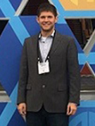 We are proud to introduce the First-Place Winner of the American Association of Physics in Medicine (AAPM) John R. Cameron Young Investigators Competition as our very own James Scheuerman, Research Assistant in the Department of Radiology Digital Radiological Imaging Laboratory (DRIL). The DRIL is led by Wei Zhao, PhD, Professor of Radiology. The award was for his research project titled; “Low-Dose Imaging with Avalanche Amorphous Selenium Flat-Panel Imager.” James’ research also received the Physics of Medical Imaging Paper Award (3rd place) the 2016 SPIE Medical Imaging Conference in California. This extraordinary award, and contribution to Radiological Imaging, exemplify the highest standards and research activity here in the Department of Radiology that help to promote and expand the boundaries of excellence in patient care.
Read more about the award.
To read more about Mr. Scheuerman, Dr. Wei Zhao and this research, please visit these links:
https://www.researchgate.net/profile/James_Scheuermann
http://www.mil.sunysb.edu/dril/index.html
http://www.aapm.org/meetings/2016AM/PRAbs.asp?mid=115&aid=32431
Grant Approval for Dr. Huang
August 2016
 Chuang Huang, PhD, has had his NARSAD Young Investigator Grant approved by the Brain & Behavior Foundation.
Dr. Fisher to Assume the Responsibilities of President of the Long Island Radiologic Society
August 2016
Dr. Paul Fisher recently assumed the responsibilities of President of the Long Island Radiologic Society. The LIRS has sponsored state of the art CME presentations and Courses for member radiologists in Nassau and Suffolk counties.
Dr. Fisher Obtains Multiyear Research Collaboration which Includes a State of the Art Mammography Unit
August 2016
Dr. Fisher has an ongoing multiyear research collaboration with Siemens Corporation ( Germany). A state-of-the-art Contrast Enhanced Dual Energy tomography mammography unit, (CEDET) of which there are only two prototypes currently existing (one here at Stony Brook, and one in Austria), is undergoing a clinical trail with selected patients from the Carol Baldwin Breast Care Center.
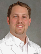
New Physician to Join the Department of Radiology
July 18, 2016
 Dr. Masha Hoshmand Kochi has joined the Department of Radiology as an Assistant Professor of Clinical Radiology. She graduated from Stony Brook School of Medicine with her Medical Doctorate. She went on to do her internship in Internal Medicine at Winthrop University Hospital, her residency here at Stony Brook University Medical Center in Diagnostic Radiology, and her fellowship at New York Presbyterian Medical Center in Body/MRI Imaging. We are very excited to welcome her to the department, along with all her experience and knowledge she will bring.
CTC Research to Receive Continued Funding
July 5, 2016
Professor Jerome Liang's, PhD, research was recently awarded the 4th term of R01 funding from the National Cancer Institute (NCI) of the NIH (National Institutes of Health), to further advance the CTC paradigm to not only detect polyps but also to differentiate malignant (adenomatous) polyps from non-malignant (hyperplastic) ones as a cost-effective diagnostic screening method.
Research on Computed Tomography Colonography (CTC), also known as Virtual Colonoscopy (VC), began over 20 years ago. The research at Stony Brook University have several technical innovations, for example, oral tagging the colonic materials, virtual cleansing the colon lumen, accurate segmenting the colon mucosa and detail-preserved processing of the segmented mucosa for visualization and computer-aided detection of polyps as small as 5 mm. This research is to lead CTC to be accepted as a clinical option recommended by the U.S. Preventive Services Task Force for screening of early colorectal cancer.
To learn more about Computed Tomography Colonography Research, please click the links below
http://technology.newscientist.com/channel/tech/dn14153-virtual-colonic-irrigation-gives-clear-view-of-cancers.html
http://www.spectrum.ieee.org:80/oct08/6791
http://www.uspreventiveservicestaskforce.org/Page/Document/RecommendationStatementFinal/colorectal-cancer-screening2#pod4
Conference Presentation by Dr. Fisher
July 2016
Dr. Paul Fisher is the co-Director, and featured lecturer, at the Second Annual “Which X-ray Is Best” conference, in NYC in July 2016.
A Triumph for CT Colonography - CTC to Gain Insurance Coverage
June 15, 2016
On June 15, 2016 the US Preventive Services Task Force (USPSTF) released the final recommendation statement for all colorectal cancer screening. This turned out to be great news for CT Colonography (CTC). Colorectal cancer screening was assigned an "A" grade in 50- to 75-year-old patients and the list of recognized screening tests to be performed every 5 years now includes CTC. An "A" rating means that the USPSTF recommends the service and that there is high certainty that the net benefit is substantial. The Affordable Care Act requires that private insurance plans now cover CTC without any co-pay. This also puts screening CTC in a strong position to obtain reimbursement from the Centers for Medicaid and Medicare Services (CMS). CTC was first introduced at a Society of Gastrointestinal Radiology (SGR) meeting in 1994. Since then, CTC advocates, many who are SAR members, have worked tirelessly for over a decade in their pursuit of national coverage of screening CTC. Members of the ACR Colon Cancer Committee include Dr. Matthew Barish of Stony Brook Medicine. The impact of this achievement will significantly change the way we screen for colorectal cancer in this country.