Clinical MRI
Stony Brook University is the birthplace of the MRI. The Department of Radiology have multiple state-of-the-art human and small-animal MRI scanners dedicated to research. Our faculty in MRI research covers expertise in novel MRI data acquisition technologies and advanced image analysis for both clinical and preclinical imaging applications.
Clinical MRI Research
Both scanners include adequate peripheral equipment including a projector connected to a PC for visual stimulation using E-Prime, a response pad used to record the subject response during the fMRI tasks. Prisma scanner also has an MRI-compatible eye-tracking system, an MRI-compatible TMS coil for noninvasive transcranial magnetic stimulation of the brain, and a BIOPAC physiological monitoring system for pulse, respiration, and GSR measurements.
Imaging capabilities: Both MRI scanners are supported by advanced MRI clinical protocols for state-of-the-art high spatial resolution images for detecting morphological and anatomical abnormalities in brain, spinal cord, chest, abdominal, thigh, knee and foot. We also equipped advanced protocols for non-invasive imaging of diffusion (DTI), perfusion (ASL) and metabolism (MRS and CSI).Expertise: MRI scientists within Department of Radiology have extensive experiences in novel MR acquisition and image analysis technologies. Novel MRI acquisition technologies include imaging brain perfusion (CBF) and oxidative metabolism (CMRO2), blood-brain-barrier disruption, and injuries of MSK including tendon, cartilage and spinal cord disk; as well as MR spectroscopy for assessing in-vivo concentration of brain metabolites such as NAA, Cho, PCr, GABA, Glutathione and glutamine/glutamate. On the image analysis front, besides traditional image analysis techniques (such as tractography, connectivity analysis, volumetric analysis, 3D visualization), we also provided state-of-the-art Radiomics and Artificial Intelligence-based analyses. We would like to establish and support collaborative research projects with both clinical and research investigators inside and outside of Stony Brook campus. We can offer consultation and some pilot MRI scanner time for promising projects.
Scanner Rate:
| Prisma $625/hr | https://you.stonybrook.edu/scancenter/ |
| Biograph mMR: | (with PET) $1,394/hr (without PET) Hospital internal rate depending on the anatomy Raw data (other than DICOM) transfer for mMR: $150/scan |
Contacts:
Discussion with clinician input: Lev Bangiyev, MD
Research on PET/MRI (Siemens): Chuan Huang, Ph.D.
Research on Prisma: Xiang He, Ph.D.
Advanced image/data analysis: Haifang Li, Ph.D., Chuan Huang, Ph.D.
Preclinical MRI Research
We provide a wide variety of small animal MRI services to investigators within and outside Stony Brook University. We encourage a wide range of collaborations to explore new imaging techniques, novel imaging contrast agents, and new applications in various animal models.
Small animal 7 Tesla MRI scanner (Bruker Biospec)
This high-field system has state of the art hardware that delivers superior imaging signal-to-noise-ratio than conventional field strength, enabling anatomical, functional, and molecular MRI research at very high spatial resolution. The system provides services primarily for in vivo rodent (rats and mice) brain and body studies but can be used for ex vivo specimens and potentially other species.
Features- Paravision 6 software for structural, physiological, and functional MRI, and spectroscopy
- Avance III HD hardware
- The gradient capability is 65G/cm with a 12-cm diameter bore size
- A micro-imaging gradient for mice is available, with 100G/cm and a 6-cm bore
- RF amplifiers for standard 1H MRI and for MRI of other nuclides (e.g. 19F, 2H)
- An assortment of vendor and custom-made RF coils
- Holders with support for rat and mouse experiments
- Equipment for anesthesia, physiological monitoring, and stimulus presentation
Our MRI expertise includes
- T1, T2, and spin-density structural MRI; Diffusion-tensor imaging (DTI); Functional MRI (fMRI); Blood flow MRI; Dynamic contrast enhanced MRI (DCE); Fat imaging; MR spectroscopy (MRS)
Current and previous projects
- We have expertise in neuroimaging and cancer imaging in various organs. Examples of several MRI applications that have been conducted at the Preclinical MRI Center are listed. If you are interested in other applications, please contact us to discuss.
Cancer imaging
- Brain cancer:
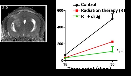 In a mouse model of glioblastoma multiforme (GBM), we have used MRI to longitudinally monitor brain tumor volume before and after a novel combination treatment of radiation therapy and an anticancer drug. We found that the novel combination therapy was more effective in reducing tumor burden and yielded better survival compared to non-treated or radiation-only. (This project is in collaboration with Dr. Samuel Ryu in the Department of Radiation Oncology).
- We have used MRI to detect brain metastases of breast cancer cells labeled with an MRI contrast agent and injected in mice. Dynamic contrast enhanced MRI was also used to detect regions of blood brain barrier leakage. (This project is in collaboration with Dr. Jun Lin in the Department of Anesthesiology).
 Pancreatic cancer:
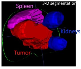
The lack of effective treatments and intrinsic chemoresistance to first-line therapeutic agents underlie the low survival of patients with pancreatic ductal adenocarcinoma (PDAC). Using MRI and MRS in different subtypes of a PDAC mouse model, we detected differences in tumor volume and in several metabolites, including the glycolytic activity indicator, lactate. (This project is in collaboration with Dr. Kenneth Shroyer in the Department of Pathology).
- Liver cancer: In a mouse model of hepatocellular carcinoma (HCC), we have used MRI to longitudinally measure tumor volume pre- and post-treatment, which reduced tumor size and led to complete tumor disappearance in a few individuals. Treatment was also associated with doubling the survival time. (This project is in collaboration with Dr. Ute Moll in the Department of Pathology).
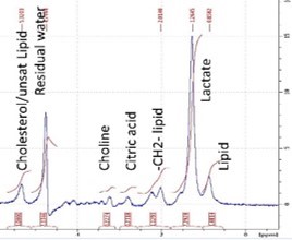 Breast cancer: We have used MR spectroscopy to measure lactate metabolism in breast cancer tumors in mice 18 days after injection of cancer cells into the abdominal mammary glands.
Neuroimaging
Stroke: We have used MRI in a rat stroke model to measure cerebral blood flow deficits, diffusion MRI to detect necrotic lesions, and T2-mapping to measure edema. We have used these methods to non-invasively investigate the efficacy of novel stroke therapeutics, such as hydrogen water. We have also used also used forepaw stimulation and hypercapnic inhalation to assess neurovascular dysfunction in stroke. (This project is in collaboration with Dr. Dennis Choi in the Department of Neurology).
Novel drug testing: We have used functional MRI to detect changes in brain functional patterns caused by administration of a novel drug. (This project is in collaboration with Dr. Alfredo Fontanini in the Department of Neurobiology and Behavior).
Olfactory system: The olfactory system may be an early site of dysfunction in many neurodegenerative diseases. We have implemented functional MRI of olfactory stimulation, diffusion tensor MRI to measure structural connectivity in the olfactory network, and manganese enhanced MRI to measure axonal transport in the olfactory system.
Rates
| Assisted |
External, Assisted |
Self-operated* |
After hours, Self-operated |
Data analysis |
| $350/hr | $450/hr | $200/hr | $100/hr | $50/hr |
*Operators must be trained and approved by Center staff. Due to the extensive time needed for training, we will only consider training users in special circumstances involving large projects.
Contacts
Research discussion: Eric Muir, PhD, eric.muir@stonybrookmedicine.edu
Implementation of scan: Zhao (John) Jiang, PhD, MD, zhao.jiang@stonybrookmedicine.edu
Website: https://renaissance.stonybrookmedicine.edu/radiology/research/centers
iLab website: https://cores.stonybrookmedicine.edu/service_center/show_external/5034