Supplementary Information for Apollo Procedure article by Stephen Probst, MD
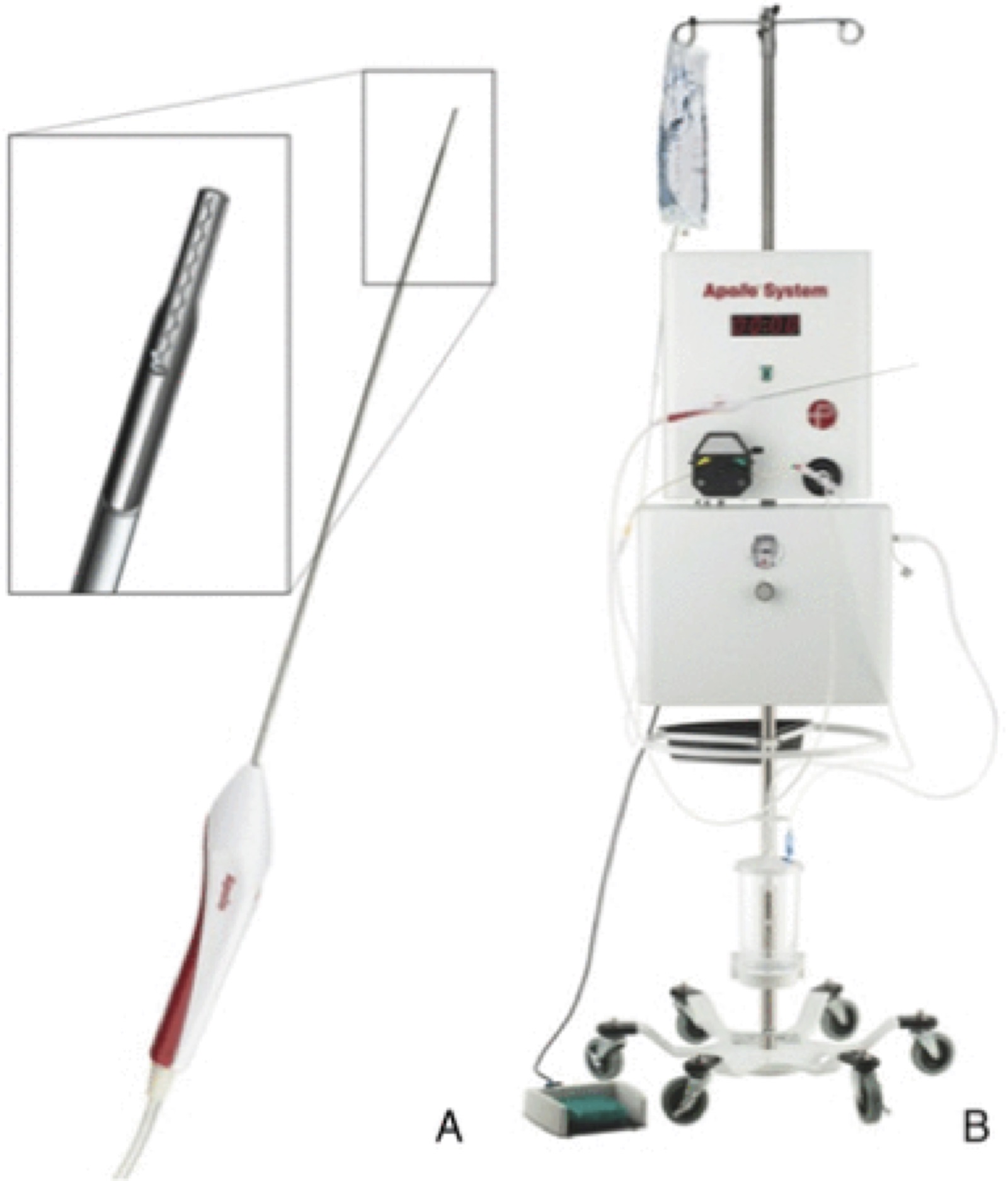
The Apollo system is composed of an aspiration–irrigation system, which can be attached via flexible tubing to the Apollo wand. The wand (A) can be accommodated by an 8F vascular sheath. The 2.6 mm wand houses an internal agitator wire that macerates clot material to maintain patency of the system during aspiration. The wand is attached to the freestanding aspiration–irrigation system (B). The aspiration–irrigation system (B) provides the capability for aspiration and saline irrigation and transmits vibrational energy to the internal agitator element within the wand.
References
1. Qureshi AI, Tuhrim S, Broderick JP, et al. Spontaneous intracerebral hemorrhage. N Engl J Med 2001;344:1450–60
2. Arima H, Wang JG, Huang Y, et al. Significance of perihematomal edema in acute intracerebral hemorrhage: The interact trial. Neurology 2009;73:1963–8.
3. Gebel JM Jr., Jauch EC, Brott TG, et al. Natural history of perihematomal edema in patients with hyperacute spontaneous intracerebral hemorrhage. Stroke 2002;33:2631
4. Mendelow AD, Gregson BA, Rowan EN, et al. Early surgery versus initial conservative treatment in patients with spontaneous supratentorial lobar intracerebral haematomas (STICH II): a randomised trial Lancet 2013; 382:397–408.
5. Teernstra OP, Evers SM, Lodder J, et al. Stereotactic treatment of intracerebral hematoma by means of a plasminogen activator: A multicenter randomized controlled trial (SICHPA). Stroke 2003;34:968–74
6. Mould WA, Carhuapoma JR, Muschelli J, et al. Minimally invasive surgery plus recombinant tissue-type plasminogen activator for intracerebral hemorrhage evacuation decreases perihematomal edema. Stroke 2013;44:627–34.
7. Hanley DF. 365-day outcome and cost model. Mistie II: a phase II proof-of-concept trial. Int Stroke Conf 2013.
8. Fiorella D, Gutman F, Woo H. Minimally invasive evacuation of parenchymal and ventricular hemorrhage using the Apollo system with simultaneous neuronavigation, neuroendoscopy and active monitoring with cone beam CT J NeuroIntervent Surg 2015;7:752-757